
Science
European Foulbrood (EFB) is a brood disease caused by the gram-positive, non-spore forming bacterium Melissococcus plutonius. Severe cases of EFB negatively impact brood population and honey production. EFB is common in the spring and early summer when populations are reaching their peak and during times of stress from lack of protein, inadequate numbers of nurse bees to feed larvae, and inability for young bees to feed enough royal jelly to larvae (Alippi, 1999).
Pathogenesis of EFB typically begins when food contaminated with M. plutonius is ingested by uncapped larvae less than 48 hours old but may also affect capped larvae in severe cases masking as AFB. M. plutonius competes for nutrients in the larval gut, eventually destroying the peritrophic membrane and intestinal epithelium (Alippi, 1999). Bacterial competition for food increases the appetite of the larvae. In strong hives, nurse bees will eliminate larvae with abnormal appetites (Alippi, 1999). EFB progresses when there is an abundance of nurse bees feeding and not removing EFB infected larvae (Alippi, 1999).
EFB symptoms may disappear when a nectar flow begins as nurse bees shift priorities from feeding brood to foraging. At this point, infected larvae will begin producing signs of EFB as they starve and die. If the decaying larvae are removed, the disease does not progress in severity and may resolve. The longer decaying EFB infected larvae remain in the hive, the more likely secondary infections are to occur. Secondary bacterial infections may exacerbate EFB and exhibit the odor and consistency of AFB. EFB larvae who survive through pupation will leave behind bacterial infected feces in the cell capable of reintroducing EFB to the hive for years to come (Alippi, 1999). Food absorption and development are compromised in infected survivors due to bacterial interference with these processes.
Secondary infection bacterial species:
- Lactobacillus eurydice
- Non-spore forming. Commonly found in healthy adult honey bee alimentary tract and midgut of larvae. Populations increased in EFB infected larvae (Alippi, 1999).
- Paenibacillus alvei
- Gram variable, spore forming – serves as indicator species for EFB. Lives on decaying larvae (Alippi, 1999).
- Brevibacillus laterosporus
- Gram variable, spore forming – found less frequently than P. alvei (Alippi, 1999).
- Enterococcus faecalis
- Gram positive. Produces sour smell (Alippi, 1999).
- Paenibacillus apiaries
- Gram variable, spore forming and rare (Alippi, 1999).
Possible Infection Pathways Review:
- M. plutonius infected larvae are removed before capping by nurse bees. Disease may abate.
- Larvae die from plutonius infection before being noticed by nurse bees. If infection becomes severe, secondary bacterial organisms may quickly colonize producing more severe symptoms that mask AFB.
- Infected survivors may show signs of developmental abnormalities and will leave traces of M. plutonius infection behind in the cell.
Transmission:
EFB commonly spreads to other hives through robbing bees or transfer of contaminated equipment. EFB organisms are capable of overwintering on hive equipment (Alippi, 1999).
Signs and Symptoms
- Spotty brood pattern usually with uncapped dead larvae present
- Discolored and/or punctured cell cappings
- Larvae in abnormal, twisted positions in comparison to C-shape in healthy larvae
- Discolored off-white, yellow or brown larvae
- Loosely attached, rubbery brown scales caused by Paenibacillus alvei
- Sour odor in advanced stages where secondary bacterial infection is present from Enterococcus faecalis
- Watery consistency compared to ropiness of AFB when “ropiness test” is performed
Diagnosis:
Newly deceased larvae may be dissected on a microscope slide to reveal the midgut. Look for transparent peritrophic membrane filled with chalky-white clumps (Alippi, 1999).
Reach out to local or state apiarist agencies for diagnostic testing or assistance checking hives for disease. EFB testing kits are also available for purchase from a variety of beekeeping supply distributors.
Samples may be sent for diagnostic testing to USDA Beltsville Bee Research Laboratory. Follow the protocol for sending samples as provided on their website.
Prevention through
Proper Nutrition
Probiotics
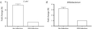
EFB can occur during stressful times in the spring, fall, dearth periods and migratory movements. Figure 1. shows a four-fold decrease in S. alvi and two-fold decrease in Bifidobacterium in the honeybee gut when an EFB infection is present. It is therefore important to support the health of the hive by supplementing with probiotics to strengthen the native immune system of the honey bees. Supplementing with a probiotic supplement like SuperDFM – Honey Bee by Strong Microbials Inc. is the perfect way to incorporate key microbial components needed to aid in the defense against pathogens like M. plutonius. The blend of commensal bacteria found in SuperDFM – Honey Bee has been tested by commercial beekeepers and proven to suppress EFB when used as recommended (four times a year).
Microbes found in SuperDFM – Honey Bee mimic the honeybee’s natural gut microbiota.
- Commensal strains of lactic acid bacteria aid in honey production, fight pathogens, maintain healthy immunity and restore microflora after antibiotic treatment.
- Spore forming bacilli create a beneficial environmental for the lactic acid bacteria, fight pathogenic yeasts and molds and reduce environmental stress.
- Enzymes like amylase, protease, beta-glucanase and cellulase help with nutrient assimilation.
- Yeasts increase fiber digestion, synthesize B-vitamins, and prevent the adhesion of pathogens to the intestinal wall.
Protein
Adequate sources of carbohydrates, proteins and lipids are honey bees first line of defense against pathogens. Adequate pollen resources during brood rearing can help avoid bacterial diseases like EFB. Pollen supplementation should not be considered a last-minute approach but should be supplemented regularly as it takes some time for the proteins and lipids to show beneficial effects in the hive. AP 23- Microbials by Dadant offers a high-quality pollen substitute with built in probiotics to promote intestinal health.
Treatment Options
- Requeening to break the brood cycle and allow nurse bees to clear away infected larvae
- Limit colony stress during dearth and migratory periods
- Avoid protein shortages during periods of increased brood development by supporting hive nutrition using pollen substitutes like AP 23- Microbials
- Treat hives four times a year with a probiotic supplement like SuperDFM – Honeybee to enhance native microbiota and suppress adherence of pathogenic bacteria to alimentary track
- Avoid treating with antibiotics which allow infected larvae to survive rather than being culled from the hive
References
Alippi, Adriana. (1999). Bacterial diseases. International Center for Advanced Mediterranean Agronomic Studies. p. 31-59.
Guo, Jun, Wu, Jie, Chen, Yanping, Evans, Jay, Dai, Rongguo, Luo, Wenhua, Li, Jilian. (2015). Characterization of gut bacteria at different developmental stages of Asian honey bees, Apis cerana. Journal of Invertebrate Pathology: Volume 127, 110-114.
1 thought on “Signs, Symptoms and Science – European Foulbrood”